
modern technology for the benefit of the patient
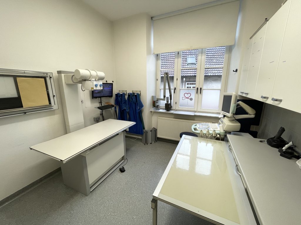
Digital X-ray
In radiology, digital x-rays are x-ray procedures in which the images are digitized either directly or via a detour using an imaging plate. The big difference to classic X-ray film is the improved possibility of post-processing, integration into the workflow of a veterinary practice or clinic and the usually lower radiation exposure.
Like conventional X-ray images, digital X-ray images are based on the projection of X-rays from an X-ray tube. The images are obtained either by an electronic detector or by subsequently scanning an X-ray imaging plate. After recording, various options for digital image processing are available.
Digital X-rays have great advantages in the process. In the past, you had to develop the x-ray image after taking the picture. Thanks to digital radiology, the images are immediately available on every PC in the system, in contrast to normal x-ray images several times. The images can be digitally post-processed at the workstation, saving you from having to take repeat shots. X-ray images can no longer be lost. Chemicals are no longer needed for film development. Another advantage is that only minimal space is required for storing the X-ray images, which must be archived for ten years. In addition, the material costs are sometimes lower because a film is not required for every recording.
We operate a digital X-ray system in the form of a fixed detector system in which the image file is generated in a similar way to a digital camera and sent to the diagnosis station.
X-rays of the chest and abdominal cavities are most commonly taken. In the event of changes, they give rise to further examinations such as ultrasound, endoscopy or laboratory diagnostics. Common findings in the chest cavity area include heart enlargement, lung opacities, increased circumference and fluid accumulation. X-rays of the abdominal cavity show organs such as the liver, spleen, kidneys, bladder, prostate, etc. If these organs increase in size, an ultrasound examination is often necessary to show the internal structure of the organs. Using so-called contrast passages, foreign bodies or other causes of obstruction in the stomach or intestinal tract can be identified. In orthopedics, x-rays are the central diagnostic tool for practically all issues. They are used to detect fractures, joint arthrosis or bone tumors, etc.
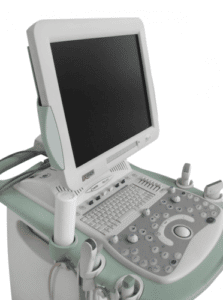
Modern ultrasound
We work with a modern, high-resolution MyLab 70, which also allows cardiac ultrasound examinations
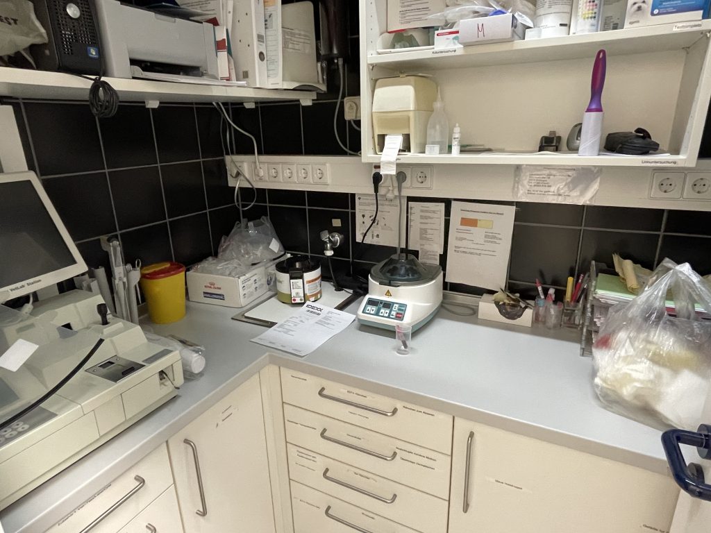
In-house laboratory
Depending on the urgency, same-day results are also possible using our in-house laboratory. We have various devices from Idexx for this purpose.